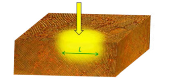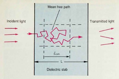
A new spectroscopic technique for turbid media is known as Diffusing Wave Spectroscopy. This enables measurement of structural and temporal properties of an opaque medium in which light undergoes a multiple scattering path. An application of this method is in diagnostic medical imaging [Journal of Biomedical Optics 1 (2), 180 (1996)]. Using multiple scattering of near-infrared light, it is possible to image brain and breast tumors non-invasively and inexpensively. Unlike X-rays that use very short wavelength radiation and magnetic resonance imaging that uses very long wavelength radiation, near IR light is an intermediate frequency probe which is sensitive to metabolic processes such as the local oxygenation level of hemoglobin. Unlike earlier methods based on the diffusion theory of light in tissue that provides resolution on the scale of a millimeter, Diffusing Wave Spectroscopy offers the possibility of resolution on the scale of 10 microns. Rather than tracking only the intensity fluctuations of diffusing light due to an inhomogeneity within a turbid medium, this new technique tracks changes in the optical Wigner Coherence function. This holds the possibility of early detection of tumors during the early metabolic stages of formation before structural damage takes place.
Related to the theory of multiple light scattering in disordered media is the phenomenon of random lasing. Our theoretical models describe (i) how this effect occurs in a photon diffusion model [Physical Review A 54, 3642 (1996) Theory of Lasing in a Multiple Scattering Medium] and (ii) the long-sought-after quantum statistical properties of light emission from the random laser [Physical Review E 69, 046603 (2004) Theory of Photon Statistics and Optical Coherence in a Multiple-Scattering Random Laser Medium].