Introduction
While this report is about the origins of the electron microscope and electron microscopy in North America at the University of Toronto, it is hoped that it will also reveal some feeling for the life of a graduate student in Physics at the University during the years of World War II and the waning years of the Great Depression, when the first electron microscope in North America was constructed and developed in the Department of Physics. I did not reach Toronto until the autumn of 1939. Consequently, what I report for the history of those years before 1939, is based upon conversations I have had with those who were there and my re-reading of the few available accounts for those years (1, 2, 3), including my own (4, 5, 6, 7, 8).
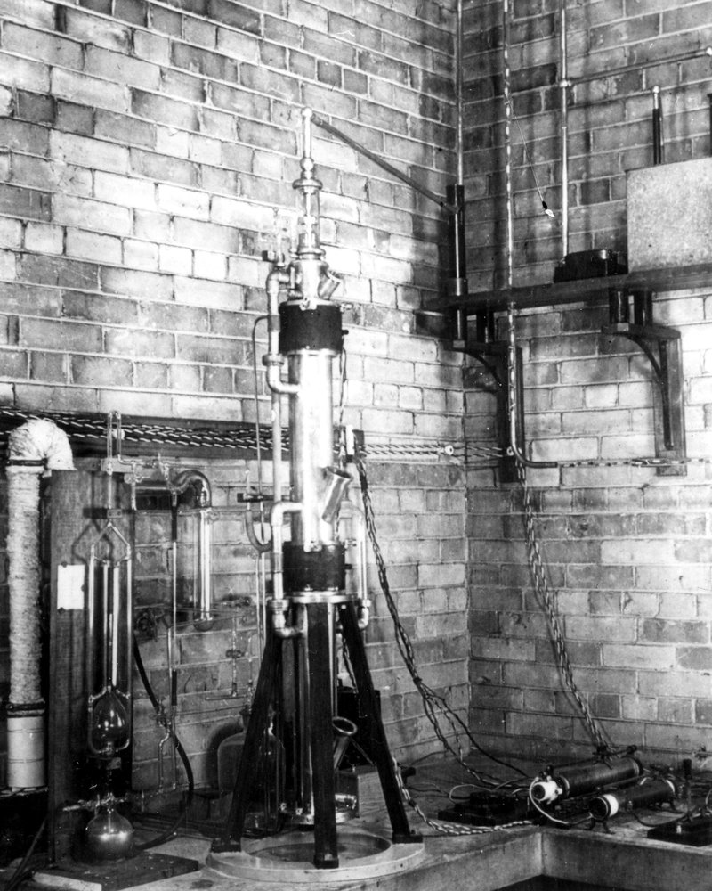
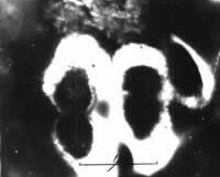
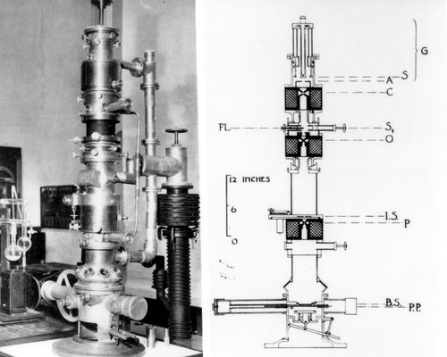
Fig. 1a: The 1938 Toronto model
Many important scientific achievements have originated from the Physics Department of the University of Toronto but in the long history of the Department I would wonder if any have been of more significance to Society as well as to science than the building of the first transmission magnetic electron microscope by two graduate students, James Hillier and Albert Prebus in April of 1938, under the direction of the Department Chairman, Professor E.F. Burton. At first the electron microscope was a subject for instrument development and for physicists but its invention quickly became a turning point for science itself. As appropriate specimen preparation techniques were developed, it became the means of relating morphology and function in controlled experiments, and the impressive modern developments, which we are all familiar with, in biology, medicine and material sciences became possible. For the first time, investigators were able to "see" directly into "inner space" that realm of the very small, below the then limit of the optical microscope, where most, if not all phenomena important in those fields find their explanation.
Immediately it was activated, the instrument produced consistently good electron micrographs, with resolution better than 140 Angstrom units (Å) and magnification of 20,000X. The microscope worked well, if inconveniently by modern standards, for six years with only minor technical improvements and without a serious major overhaul, evidence for the skill and care exercised in its construction and the intelligence applied in its design. It was the prototype for the several instruments which soon followed, constructed by Cecil Hall in 1939 at Eastman Kodak, by Albert Prebus in 1940 at Ohio State University, for the 1940 and 1944 models built by the Toronto Physics Department in those years and for the Radio Corporation of America's series of commercial instruments which was the equipment chosen by laboratories worldwide for a generation. The building of the 1938 Toronto microscope and its immediate and successful, practical operation was a remarkable achievement. It is this story and parts of its sequel I am recounting here.
The History of the Building of the 1938 Toronto Microscopy
New ideas are never confined to one person or place and in 1937 when Hillier and Prebus were beginning their work, attempts at building electron microscopes had been going on in Europe for about 10 years. Hans Busch's 1926 paper which showed theoretically that a coaxial magnetic or electric field could be expected to focus an electron beam and Louis deBroglie's earlier hypothesis that electrons possess wave properties, suggested to physicists that an electron microscope was not only possible but that it might produce resolution far superior to that of an optical microscope. The basic principles of electron lenses had already been worked out in Europe and Ernst Ruska and his colleagues at the Berlin Technische Hochschule had developed the essential design of pole-pieces to concentrate a magnetic field. Ruska himself had already constructed in 1933 his early model of a two-stage transmission instrument with which he had achieved resolution only slightly better than that expected from a light microscope. By 1933, Marton in Belgium and Martin, Whelpton and Parnum in England had also built instruments, and developments were proceeding likewise in Japan and in the United States but with less success. Of all these attempts, only Ruska was able to produce micrographs and these did not demonstrate satisfactory resolution. As Hillier noted in his M.A. thesis (9), describing work going on elsewhere, "reports carried more details of problems than of their solutions." In general, the lenses demonstrated chronic distortion and astigmatism and the few published micrographs were badly blurred. The specimens seemed to have been damaged by the heat of the beam and many, as yet unknown factors were affecting images adversely. Leading microscopists, particularly in the United States, were very skeptical of the possibility of an electron microscope and had labeled the project of creating one as "impossible" and the few published electron micrographs as "fakes." It is with this background, that the success of the two Toronto graduate students, directed by Professor Burton, was so unforeseen and so remarkable.

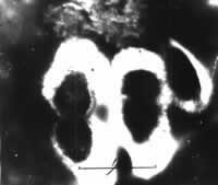
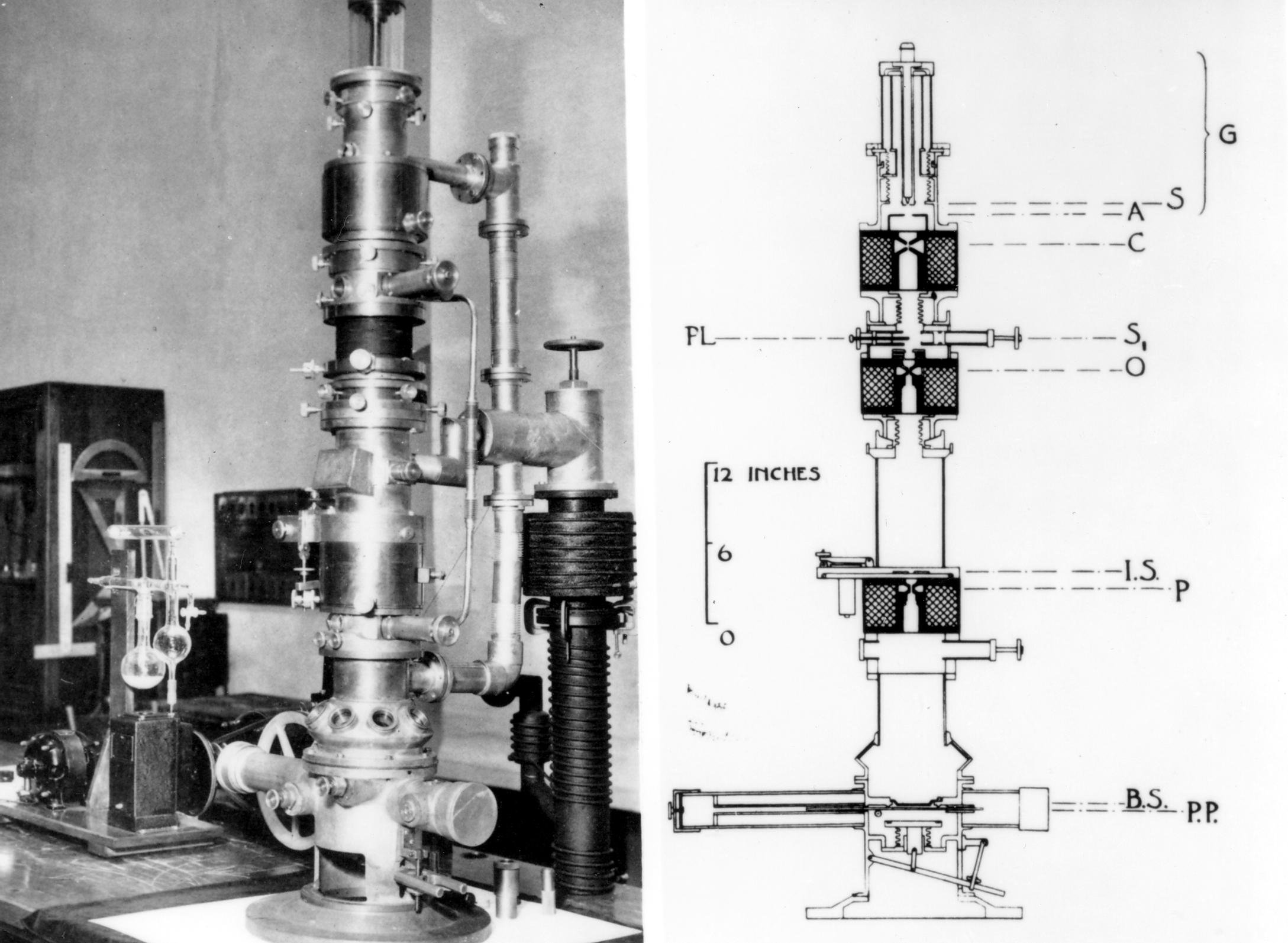
Fig. 1b: The 1938 column with the revised camera and its cross-sections.
The moving force behind the project to build a magnetic transmission electron microscope at the University of Toronto was Professor E.F. Burton, who had succeeded Sir John Cunningham McLennan as Chairman of the Department of Physics in 1932. Burton had graduated from Toronto with honours in mathematics and physics and had worked for two years as a Demonstrator under McLennan, before an Exhibition Scholarship took him to Cambridge and the Cavendish Laboratory, where he worked under J.J. Thomson, the recent discoverer of the electron. Several reasons can be advanced for Burtons keen desire to develop an electron microscope. At Cambridge he had earned a second B.A. for his work with colloids and he reasoned that an electron microscope would have resolution¹ sufficient for him at last to be able to see and study colloidal particles directly in solution and aerosols. Working for J.J. Thomson must have stirred his interest in electrons, and his own diabetic medical history would have sensitized him to the development of science in medicine, where an electron microscope might be expected to play a part. For whatever of several reasons, Burton was quick to recognize the potential of an electron microscope for research.
¹ [Resolution with a light microscope is given by the expression 1/3(lambda), where (lambda) is the wavelength of the radiation being used. With ultraviolet radiation, where (lambda)=3000 A, and using quartz lenses, the best resolution would therefore be about 1000 Ac, where one Ac = 10-8 cms. DeBroglie's theoretical wavelength of an electron is given by (lambda)=h/mv, where h is Planck's constant and m and v are the mass and velocity respectively of the electron. From this, it can be shown that the wavelength of an electron is equal approximately to 12.24/(square root)V x10-8 cms, and that with V=50,000 volts the theoretical resolution of an electron microscope would be about 0.02Å-, a considerable improvement over the resolution expected with an optical microscope.]
He was catalyzed to action by his friendship with Dr. Walter H. Kohl, who had a Ph.D. in Engineering Physics from the Technical University of Dresden and was employed in Toronto as a development engineer with Rogers Radio Tubes Ltd., doing pioneer work in television on problems to do with the deflection of electron beams by magnetic and electric fields. Kohl repeated in his own laboratory much of the published work coming out of Germany. He attended seminars in the Department of Physics and became acquainted with Professor Burton and Burton came to rely on Kohl for his knowledge of electronics and for his ability to translate German papers. At Burton's invitation, he gave many seminars in the Department on a variety of subjects to do with the emerging subject of electron optics. Kohl's lectures and demonstrations in electron optics certainly must have encouraged Burton to develop his own electron microscope project.²
²[For more detail on Dr. Burton and Dr. Kohl, the reader is referred to references 1 and 3 of this paper.]
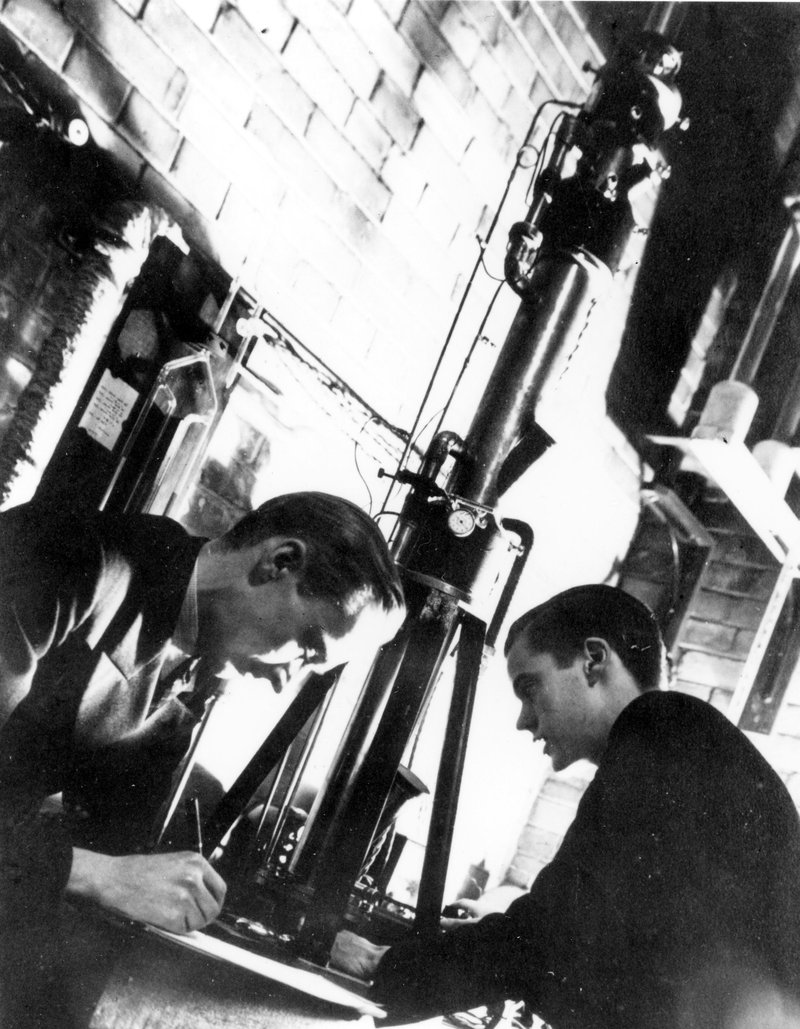
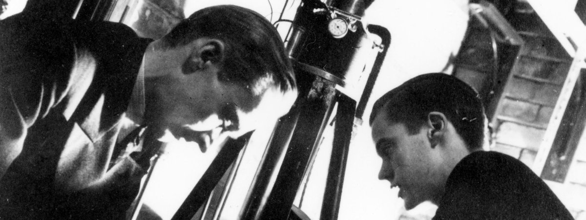
Fig. 1C: Prebus left and Hillier right, working with the microscope in 1938.
During the summer of 1935, Burton attended a conference in Berlin on the subject, Possible Areas for the Application of the Electron Microscope. In the autumn, he returned, enthusiastic, to Toronto to meet a young Cecil E. Hall, from the University of Alberta, who was expecting to do graduate work in spectroscopy. Burton was able to convince Hall, as he would later convince Prebus, to change his interests from spectroscopy to electron optics and to become his first graduate student in the new field. It is said that the general attitude of graduate students in the Department toward electron optics at that time was negative but Hall was attracted to it and agreed to construct a simple electrostatic emission type electron microscope. He built two emission microscopes during the next two years, the second with two magnetic lenses with which he took micrographs of the emitting surfaces of hot cathodes at 3000X. For this work, he received an M.A. in 1936 (10). Burton had been able to find financial support for Hall in spite of the Great Depression and the great scarcity of research moneys. The situation has a modern-day ring to it! The figure secured by Burton for Hall's second year is interesting by todays standards: 800 dollars from the N.R.C. to cover both equipment and stipend.³
³[Things had not improved much salary-wise by the autumn of 1939, when I, as a graduate student, was demonstrating Physics for a stipend of $800. for the term.]
In May of 1935, Burton turned again to N.R.C. for funds, "to attempt to take electron micrographs of substances placed in the path of an electron beam," i.e. to construct a true transmission electron microscope. Burton asked for $724.50, $250 for equipment and the balance for the investigators salary at $62.50 per month.4 On the grounds that the work could be as well done by a scholarship holder, Burtons request was refused and Hall, unfunded, was forced to leave the project. As Halls career was to show, his departure from Toronto was a huge loss for the Department and the University. Hall accepted a position at the Eastman Kodak Company in Rochester, New York., where there was interest in building an electron microscope in the United States in 1939 at Kodak. I shall not follow Halls outstanding career any further than to remark that he went to M.I.T. from Kodak and did remarkable work there in the Biology Department. At M.I.T., he completed the requirements for the Ph.D. and published his book on electron microscopy (11), still a worthwhile read for electron microscopists.
4[From these figures, it appears that Burton must have thought that Hall would be able to construct a microscope in about 7 ½ months. He wasn't far wrong. As it turned out, it took Hillier and Prebus about 4 months!]
As has been pointed out earlier, expectations for electron microscopy were viewed with extreme skepticism by many scientists. Hall has told me that optics of the electron variety was a rather "cool" subject at Toronto in the thirties when low temperature physics and spectroscopy were the "hot" topics. Dr. M.F. Crawford had been made head of spectroscopic research in 1937 and had accomplished a large body of well recognized research. He was already an authority in atomic spectroscopy, attracting many graduate students to his well equipped laboratory, including Hall and Prebus. Some of the local antipathy to Burtons electron microscope project may well have been caused by his penchant for convincing graduate students to work in electron optics rather than in the well established field of spectroscopy. Albert Prebus had come to Toronto in the autumn of 1937 fully intending to continue his interest in atomic spectra, but Burton convinced him too, to change his research interests into the new field. During the years that I knew them, I must admit that I never heard either Hall or Prebus complain that they had deserted spectroscopy for electron optics.
When Prebus arrived in Toronto, he had an M.Sc. from the University of Alberta, and Burton teamed him up with a 1937 B.A graduate in M and P from the University of Toronto, James Hillier, who had already been working since graduation on the electron microscope project. While the two are quite different in personality, with Hillier outgoing and Prebus more introspective, both are highly intelligent and diligent and were enthusiastic for the project. They worked exceptionally well as a team. Their project was to build a compound, transmission, magnetic electron microscope and to apply it to biological and colloidal materials. In order to accomplish this, in addition to their own wits, they had only Halls M.A. thesis, his emission microscopes and translated publications by Knoll, Marton and Ruska to help them.
The design, machining and construction for the column, particularly of its more sensitive components, such as the pole-pieces, were done mostly by Hillier and Prebus themselves. Prebus recalls that the shop work was done on a two-shift basis, with the day crew.5 from 8:00 a.m. to 5:00 p.m., Hillier and Prebus the night shift (without the unqualified approval of the day crew), often until 4:00 a.m. Working drawings were submitted at the end of the Christmas vacation of 1937 and active construction was begun in January of 1938. Impossible as it may seem to us now, they succeeded in assembly of the high voltage and the vacuum systems, and in construction of the column in less than four months and realized success in April of 1938 with quality micrographs at measured resolution of 140 Å, soon improved to 60 Å by use of a solid objective pole-piece, machined en bloc and designed by Prebus (12). I can witness personally that many micrographs with 20 Å or better measured resolution were taken with the 1938 model during the years that followed. Prebus has confided in me since, that he and Hillier were somewhat surprised at their initial quick success. I imagine that they must have been, but there must have also been extreme gratification and pleasant excitement for both of them.
5[The personnel of the McLennan Laboratory machine shops were very important to the develop-ment of electron microscopy at the University of Toronto. Mr. Grantley Woodward, particularly, did much of the construction of the Toronto microscopes and he acknowledged the efforts of his colleague, Mr. Frank Shepherd, who, like Grant, worked on both the 1938 and 1940 models. Grant was mainly responsible later for the machining and construction of the 1944 model. For his technical contributions to early electron microscopy, Mr. Woodward was awarded an honorary membership in the Electron Microscopy Society of America in 1978. He worked with each of us and much of his time was spent deciphering our amateur engineering drawings, on which we left our spurts of genius. In the basement of the McLennan Laboratory there was a true genius, a blower of glass, Harold Chappelle, who blew our first mercury and diffusion vacuum pumps, and whose talented hands could produce any object of glass, however complicated, for us or others at the Laboratory.]
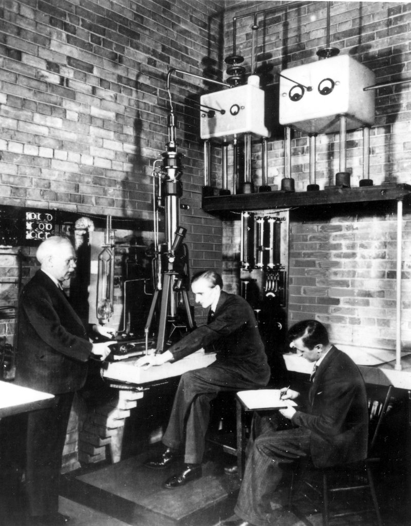
Fig. 1D: The 1938 microscope with the revised camera, Prebus at the microscope, Professor Burton left and Ladd sitting right, 1940.
The successful outcome of the project was reported by Burton to the University in his very casual way, "Mr. James Hillier, assistant demonstrator and Mr. Albert Prebus, holder of a studentship from the National Research Council have continued the work of perfecting the electron microscope and have succeeded in taking many photographs of submicroscopic structures up to a primary magnification of 30,000 times". The first scientific report was submitted to the Canadian Journal of Research in January 1939, just one clear year after work had been begun, and appeared in April 1939 (13). A short note was published in Physical Review in December 1939 (14) and other reports, describing the work, appeared in 1939 (15) and 1940 (16). Some of the first micrographs were reproduced on December 17, 1938 in the popular magazine Saturday Night with a descriptive text by Burton, and a second popular article appeared in MacLeans magazine on April 1, 1939 authored by the journalist R.E. Beamish. The book by Burton and Kohl (17) which described the 1938 microscope appeared in 1942, with its second edition, describing the 1944 model, in 1946.
I happened to read the MacLean's story in the spring of 1939, when I was in my graduating. year in Mathematics and Physics at McMaster University in Hamilton, Ontario. I was intrigued by the description of a new type of microscope which used a magnetic field instead of glass as a lens, and electrons instead of light as radiation, and wishing, in any case, to continue with University life, I wrote to Professor Burton asking if I could be considered for a situation as a graduate student in Physics under his direction. To my delight, and somewhat to my surprise, Dr. Burton accepted my application and offered me a fellowship in his laboratory. Thus, by reason of reading the MacLean's article, the generosity of Professor Burton and my own good fortune were my feet placed on the pleasant and rewarding paths of electron microscopy.
William A. Ladd, a 1939 graduate in Mathematics and Physics at the University of Toronto, whose name is familiar to electron microscopists as the founder of the Ladd Research Instruments Inc., also joined the electron microscope team in the Fall of 1939. He was assigned to Hillier and I to Prebus as our mentors. Ladds activity took him primarily into work with Hillier and Prebus as the second, or 1940 model electron microscope was built. Funds for this were provided by the Colombian Carbon Company of Brooklyn, New York, whose far-sighted research director, Dr. W.B. Wiegand, a graduate in physics of the University of Toronto and a close friend of Burton, had realized early the potential of the microscope for determining the particle sizes and other properties of carbon blacks. Ladd left the laboratory after receiving his M.A. (18) in 1940 and went with the 1940 microscope to Brooklyn.
My M.A. studies were related to basic electron optics with Prebus and my M.A. thesis (19) was entitled. "The Measurement of the Magnetic Field Along the Axis of an Electron Lens". I also used the 1938 microscope with biological specimens, helped to improve its operation and received the M.A. degree in 1940. I stayed on at Toronto to receive my Ph.D. in 1943 (20), after applying the microscope to a wide range of specimens from both biological and material science and developing the designs of the third, or 1944 model Toronto microscope, which became a part of my Ph.D. thesis. In September of 1943 I joined the Physics staff of the University of British Columbia, where I stayed for two years and did no electron microscopy except to talk about it. I returned to the Toronto laboratory only for the summer of 1944 to help bring the 1944 microscope into operation. In September of 1945 I took up a new position as physicist with the Shawinigan Chemicals Ltd. in Shawinigan Falls, Quebec, where I stayed for two years doing industrial electron microscopy before taking a position in medical research in the Henry Ford Hospital in Detroit, Michigan, where I stayed.
Prebus and Hillier left Toronto in 1940 to continue their careers in the United States. Hillier who had received his M.A. in 1938 and who returned to his alma mater for his Ph.D. in 1941 (21), accepted a position at R.C.A. where he enjoyed great success, received many honours and became R.C.A. Executive Vice-President and Senior Scientist. Prebus also did extremely well, accepted a post-doctoral position at Ohio State University, later became a full Professor at O.S.U. and continued his interests in theoretical electron optics. He had received his Ph.D. degree from the University of Toronto in 1940 (22).
The "Golden Age" of electron microscopy at Toronto coincided with the years of World War II, 1939 through 1945, when many of the electron microscopy projects were related to the war effort and were classified. Consequently, few publications from those years exist. In the Department of Physics the development of the electron microscope, as an instrument, ended with the completion of the third microscope, which had involved my self with Dr. Lorne T. Newman. Dr. Newman was a University of Toronto graduate, a colloid physicist who came on staff in 1940, advised me on my Ph.D. program, and left the laboratory later to join the Manhattan Project in Oak Ridge, Tennessee. At the same time as Dr. Newman arrived, Dr. Beatrice Deacon, also a colloid physicist, and a Toronto graduate, came on board. Her work was confined exclusively to war-related quantitative studies of the filtration efficiency of a variety of fibrous materials and of the particle sizes of debilitating solid-particle aerosols, incident upon those materials, studies in which both Dr. Newman and I were also involved.
The 1938 Model Toronto Electron Microscope and its Operation
The 1938 Toronto model was about six feet tall, stood on a concrete shelf, (Fig.s 1A, B, C, D) was constructed in six sections, each joined to the other by vacuum-tight (it was hoped!) plane-lapped, vacuum-greased seals, as an upright vertical column of the six sections. This arrangement allowed horizontal, sliding adjustment of each section for alignment of optical axes and for selection of specimen areas. The vertical construction was reproduced in almost all of subsequent commercial models, with the gun at the top and the camera at the base. The Philips Co. produced an early model which lay on its side but it never caught on. With it, I always had the strange feeling that the electrons might fall under gravity and miss the photographic plate! A professorial friend of mine once observed that it would make a good examination question: how far would a 45 KV electron fall under such a circumstance? I have heard that someone did devise a reversed vertical column with the filament at it base and the camera at the top. With it, in addition to a rather dangerous location of the high voltage for the operator, the use of a step ladder to view the image also seemed a major problem. The model never caught on either!
The first or top section of the column was composed of the electron gun and the condenser lens as a unit. At first, Hillier and Prebus had tried using a cold cathode gas discharge tube as their electron course, most likely in order to provide an approximation to an ideal point source. With it there proved to be just too many problems and a hairpin tungsten filament was finally chosen, with means of tilting it and moving it horizontally in two directions, and of raising and lowering it with respect to the shield. A maximum of 45 KV was supplied to the gun by the high tension system, and the filament current by two 12-volt storage batteries in series, mounted in an insulated box, which formed a part of the high tension system. The whole gun assembly was joined rigidly to the condenser lens by a glass cylinder sealed in wax at both ends.
One of the more inconvenient characteristics of the design of the top section was met whenever a filament had to be replaced, and with poor vacuum this was a frequent occurrence. To replace a filament, the glass cylinder had first to be separated from the gun by melting the wax seal with a blowtorch. Next, a new filament had to be manufactured (as with everything else we made our own filaments), by bending 13 mm. of 6 mil. tungsten wire into a "V", with a radius of curvature at its tip of about 0.05 mm. This was then spot-welded to the electrodes of the gun and the gun returned to its position at the top of the column by remelting the wax without breaking the cylinder or burning ones hands off! The whole arrangement had to be air-tight or further flaming would be required and, oh yes, the tip of the filament had to remain centered in the hold in the cathode shield or the whole process had to be repeated. Changing a filament in 1939 bore little resemblance to the method of 1995, when a pre-centered, filament assembly, purchased from a supply house is plugged into the gun, which has not been removed from the column but merely swung back from its neoprene seal. For a long time we had no neoprene!
Such an apprenticeship for modern electron microscopists would serve no useful purpose, but it did give us an understanding and an appreciation of how an electron microscope works, which it would have been difficult to achieve otherwise, and probably too, a better appreciation of the modern microscope and the informative micrographs it produces. It is amusing now to recall, that when we achieved a micrograph which clearly demonstrated the first Fresnel diffraction maximum, we thought it a moment for cheering, not jeering, "under focused."
The second section of the column was the specimen chamber which rested immediately upon the objective lens, separated from it and from the condenser above it by a thick film of vacuum grease, which allowed horizontal movement over the objective lens of the chamber and of the specimen cartridge it carried. I should pause here to emphasize, that we also made our own vacuum grease. Hillier has written that "we used pure gum rubber, gradually stirring it into a large beaker of boiling Vaseline in order to make stopcock grease of the right consistency and sufficiently low vapour pressure." Perhaps that is the way he did it but by the time I arrived at the laboratory, this hot, tedious job had been given to the low man on the totem pole, viz. me, and the pure gum rubber had given way to elastic bands! The tacky grease, which resulted from either method held a vacuum sometimes but was one source of the "pounds" of contamination we produced operating the microscope. The other sources were oil from our own hands while handling interior components of the instrument and later oil vapour from the diffusion pump itself. Contamination in electron microscopy is the solid hydrocarbonaceous, polymeric material deposited on surfaces when an electron beam strikes hydrocarbon vapour. Of course, it was no problem to us, because contamination6 had not yet been discovered! We merely had to clean it up continually.
6[ I had reported the effect of contamination on electron microscope specimens, (23,24) but I did not use the term at that time. Hillier was shown the effect of contamination on carbon black particles when he visited the Shawinigan Laboratory in 1947, where I was using the first commercially purchased electron microscope in Canada. He said that they had noticed the effect at RCA but had not reported it. Hillier was in Shawinigan Falls to attend a symposium on electron microscopy supported by the Shawinigan Chemicals Ltd. This was the first such meeting held in Canada. Representatives from McGill University, the NRC, RCA, the University of Toronto and from the Shawinigan Company attended the meeting, (Fig. 2).]

Fig. 2: Canadians interested in electron microscopy met in the Spring of 1947 at the Shawinigan Chemicals Co. in Shawinigan Falls, Quebec. Glen Ellis is at far left in the front row. Dr. Keyes of McGill University is beside him and Professor Burton and James Hillier are beside Keyes in the front row, with the Author's head between them in the second row. Others are representatives from McGill University, the National Research Council and the staff of the Shawinigan Company.
Continuing with this description of the second section of the instrument, the specimen cartridge was supported within it on an internal platform. The cartridge contained a 1/8 inch specimen disk, coated with a thin supporting film of collodion, which was placed at the top of the cartridge, the length of the cartridge being such that the specimen was located immediately above the front focal plane of the objective. At first, the specimen disk was punched out of platinum sheet, and a single tiny hole was drilled at its center. The expensive platinum was soon replaced by less expensive copper and the weak collodion by tougher Formvar. Finding that single hole involved a tedious procedure of trial and error, sliding the object chamber back and forth over the objective until the hole was located, often with neither supporting film nor specimen still present. Eventually, someone had the brilliant idea of punching the disks from flattened screen door screens or from flattened bronze wire mesh from sieves used next door in the Chemistry Department. Suddenly, we had many collodion-covered holes bearing our specimens and our batting average improved. The specimen cartridge was inserted via a port in the side of the chamber, after air had been allowed into the column. Whenever a specimen was changed, a photographic plate introduced or removed, or an aperture taken out for flaming, vacuum was lost in the whole instrument and absorbed gases would have to be removed subse-quently by our very inadequate pumping system. As has already been indicated, the 1938 microscope worked well but inconveniently.
The important third section was the objective lens itself, which produced an image magnified 100 to 125X. Initially, its pole-piece was in three parts, with the upper and lower pieces separated by a brass spacer, which determined the size of the gap and helped to align the upper and lower pieces. With it, an inevitable misalignment occurred to limit the resolution. After the pole-piece was revised (12) measured resolution was often as good as 20 Å during the years that followed. There was a tiny set aperture in the objective, made again by drilling a small hole in an 1/8 inch disk of platinum. Every effort was made to center this aperture as it was introduced into the objective, but it was often necessary to complete the centering during operation. There being no external controls for this, it was accomplished by striking the side of the objective sharply with a rubber mallet, while observing the aperture image on the screen! Not the most acceptable of techniques, but it worked!
The image from the objective was viewed on an intermediate fluorescent screen at the lower end of the fourth section of the column. This section was a long brass tube, within which was a co-axial soft-iron or µ-metal tube to shield the beam from external magnetic or electric fields. Until this improvement in shield-ing was realized, it was advantageous to work at night, when stray fields and vibration from electric streetcars rattling along College Street, or from trucks and buses bustling along near the building were less frequent and electro-magnetic effects from a close-by, ancient elevator were not so liable to be present to affect our images. Working at night could be hazardous, as Glen Ellis discovered when walking home from the laboratory late one night along St. George Street, with his hands in his pockets. He fell off a curb and broke his collar bone. From overwork he said!
Vibration errors were also introduced by our forepump, so that it was frequently necessary to turn the pump off briefly during plate exposure. At 20,000 or 50,000X magnification, it does not take much vibration to ruin an electron micrograph! The sensitive electron image itself is the best measure of image errors from vibration, stray fields, lack of alignment, dirty apertures, lens aberrations, high voltage ripple, chromatic aberration, etc., etc. Much of the tedious work of the early electron microscopists consisted of recognizing and eliminating these disturbing factors.
The fifth section of the column was the projector lens, which magnified the objective image another 330X, to give a final magnification in the 30 to 40,000X range. We did not yet have replica gratings nor standard emulsion particles by which to determine magnification. Before these things became available, a relatively large, but still small object, whose size could be determined by optical means was compared with measurements made on the same object in the microscope. By such a method a graph of magnification versus lens current was prepared and used.
The sixth section was the camera, which contained a final viewing screen and under it the photographic plate. In the original instrument there was a single glass plate and whenever it was inserted or removed for development, air had to be allowed to enter the entire column. Hillier and Prebus had attempted to use roll film but after an enormous amount of time spent outgassing the film, it became brittle and cracked and was quite useless. Fig.'s 1A and C show the microscope with the original single plate camera, Fig.'s 1B and D show it with the revised camera for 10 x 2 inch plates and an air-lock. Neoprene had become available! After focusing the image on the screen, the operator took the micro-graph by lifting the screen, by means of a rod through a conical, vacuum-greased joint in the wall of the camera.
The original vacuum system was very slow and elementary by today's standards and was undoubtedly responsible for much of the contamination we encountered. It was not long before the mercury diffusion pump and the Hyvac forepump we inherited, were replaced by an oil diffusion pump backed by a Hypervac. The vacuum measuring equipment was a simple discharge tube and a McLeod gauge. A large percentage of our time was spent finding and correcting vacuum leaks.
Because the focal length of a magnetic electron lens is inversely proportional to the square of its field strength and directly proportional to the square of the velocity of the electrons, it is mandatory for the quality of the image, that both lens current and high voltage be kept as constant as possible. To effect this, the lens currents were supplied from well charged, 24 volt storage batteries, controlled by two rheostats in series, for coarse and fine adjustment of each lens, and the ripple in the 45 KV source was reduced to one volt in 40,000 by a resistance-capacity filter, with two standing high voltage condensers.7 A step-up transformer provided the 45,000 volts and a half-wave rectifier (Kenetron) rectified the 25 cycle power which was in vogue in Ontario at that time. The high tension system filled a room and was completely exposed to air.
7[The transformer was borrowed from a local hospital and the two condensers were borrowed from the University of Alberta. The University of Alberta had quite a stake in the Toronto electron microscope project, to which it contributed the essential two condensers and two of its more brilliant graduates, Hall and Prebus.]
It is difficult to conceive of now but a pride to remember, that I was once brave, or foolish enough to adjust an electron gun with the unshielded 45 KV voltage so close that the hairs on the back of my hands, or my youthful head were raised. The 1938 microscope was operated with a high voltage supply that was completely unprotected. One just had to remember to keep well away. Prebus had had a serious accident when he leaned against one of the condensers which, unknown to him, was charged. Frank Boswell also tells of an occasion when he experienced the effects of 45 KV. The gun and the high voltage were always sup-posed to be discharged immediately after use by means of a ground wire attached to the end of a long (30") insulator rod. Frank told me that someone borrowed the rod one day and returned it to its usual position with the wire dangling, instead of being re-attached to the ground. He later took the rod confidently in hand to use in the normal way, not knowing that the wire was free. Standing, fortunately, on a well insulated wooden stool, he suffered no permanent ill effects.
The 1938 instrument, the first practical electron built in North America which did so much to stimulate the rapid growth of electron microscopy in the world, was displayed in the early 1970's in the Ontario Science Center. Here it was presented exactly as it had been when in use in its original location in the McLennan Laboratory, on the identical cement slab, with the identical red brick wall rescued from the original McLennan Laboratory building after the Feb. 1977 fire, with the original rheostats, condensers, vacuum equipment, all arranged as they had been long ago during my student days. Even the same tall stool upon which each of us had sat to view specimens and to adjust the 'scope was there. The room was dimly lighted and the voice of the late Professor Burton could be heard from a tape, describing the instrument. It was a pleasantly eerie experience for me to enter that display room. I felt great regret in 1978, during the meetings of the International Federation of Electron Microscope Societies hosted by the Canadian Microscopical Society in Toronto, to find that this most impressive display was gone, and that the instrument was now offered only as a rather uninspiring exhibit, high up on a wall behind glass. I felt that this major Canadian achievement deserved better treatment.
Application of the 1938 Model Toronto Microscope
The greatest disadvantage we enjoyed in our time was lack of specimen preparation techniques, and it was this that made it difficult at first to convince others concerning the future of the instrument and its potential importance to future research and application. There was as yet no thin sectioning and no shadow casting, although shadow casting had been announced by Williams and Wykoff before I left Toronto and we did use it when studying lubricating greases to demonstrate the helical nature of the lime-based soaps. There was no silica replication, no carbon films, no negative staining, no cryo- nor immuno-electron microscopy. Almost all of our specimens were either dried from solution, or caught, already dry, upon collodion or Formvar film.I regret that there are so few representative early electron micrographs but very few were kept and those that I do have, have lost some quality, having been photocopied from prints discovered in the dust of my attic while preparing these texts. Hillier and Prebus used such specimens as the edge of a sharpened razor blade, carbon black particles, diatoms and viruses as their first test objects, (Fig's 3, 4).
88[It is emphasized that each of the micrographs reproduced here is a product of the 1938 microscope.]
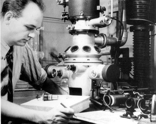
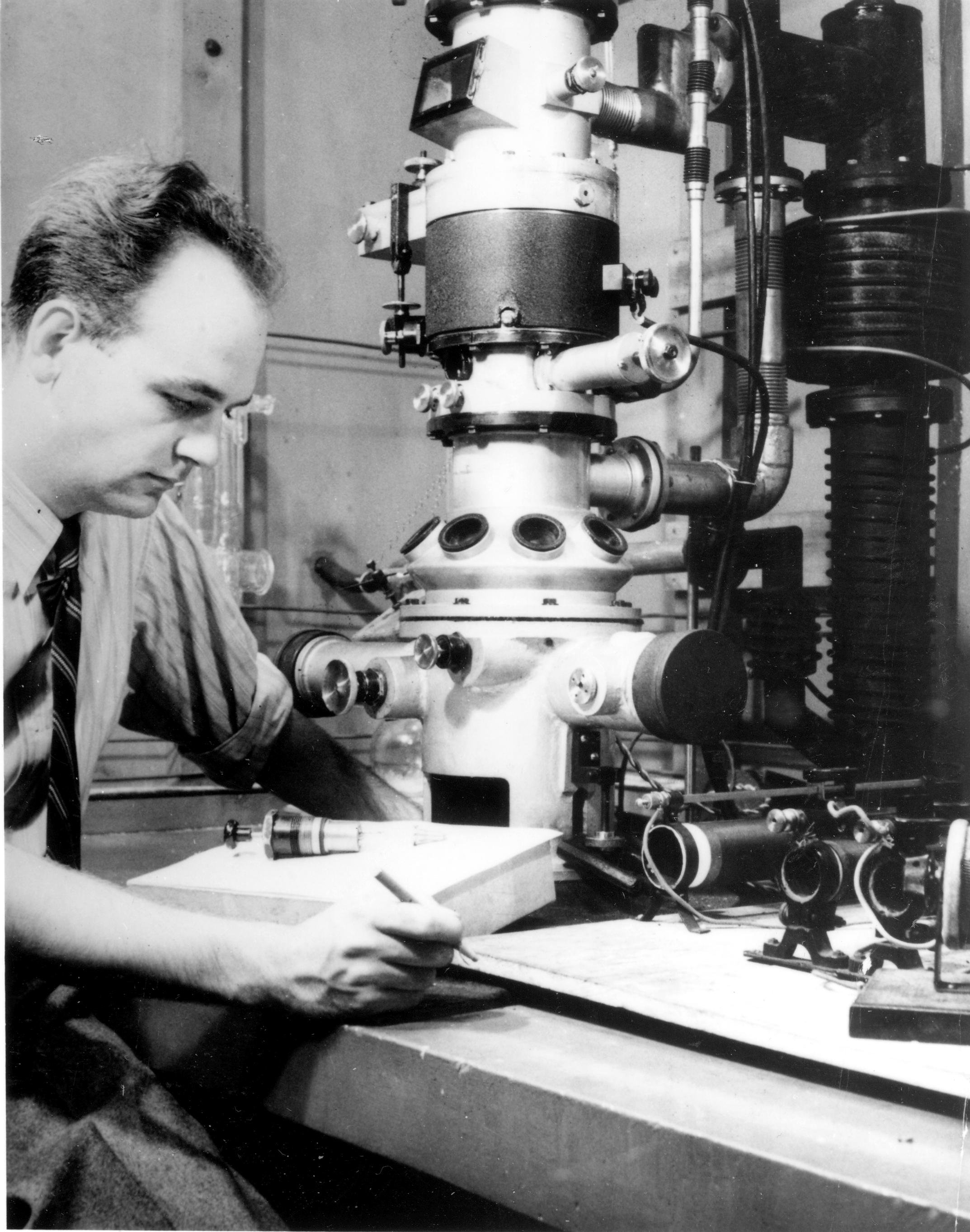
Most of the projects after 1939 were war-related and were classified. Therefore, any accumulation of data for my doctorate thesis became a problem. However, I was able to find a few unclassified subjects (25). For one thing, I began the design of the third Toronto model electron microscope, (Fig. 5), based on experience with the 1938 model. These designs became a part of my thesis. Figure 6 shows a young Watson in 1944 pondering a problem related to the specimen cartridge of the 1944 instrument.
Dr. Frank Boswell, who received his Ph.D. in electron optics in 1950 and is now a retired professor of Physics at the University of Waterloo, found the 1944 instrument of particular value for doing his high resolution diffraction and lattice studies with microcrystals (26). His work is still a classic on the subject. Quoting Boswell, "the 1944 microscope worked exceedingly well and was a godsend for my personal research because of its unique designs which provided maximum control of apertures and of the distance from specimen to photographic plate both essential for high resolution electron diffraction." In 1947 the Radio Corporation of America donated one of their commercial electron microscopes (an RCA EMU2) to the Department of Physics, recognizing the great contribution made by the University of Toronto and Professor Burton to electron microscopy. Boswell used this RCA microscope in his viral studies (27) and was able to "borrow" its high voltage system on occasion to provide improved high voltage for the 1944 model.
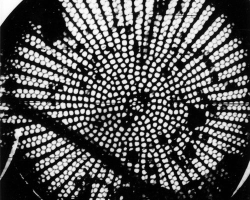
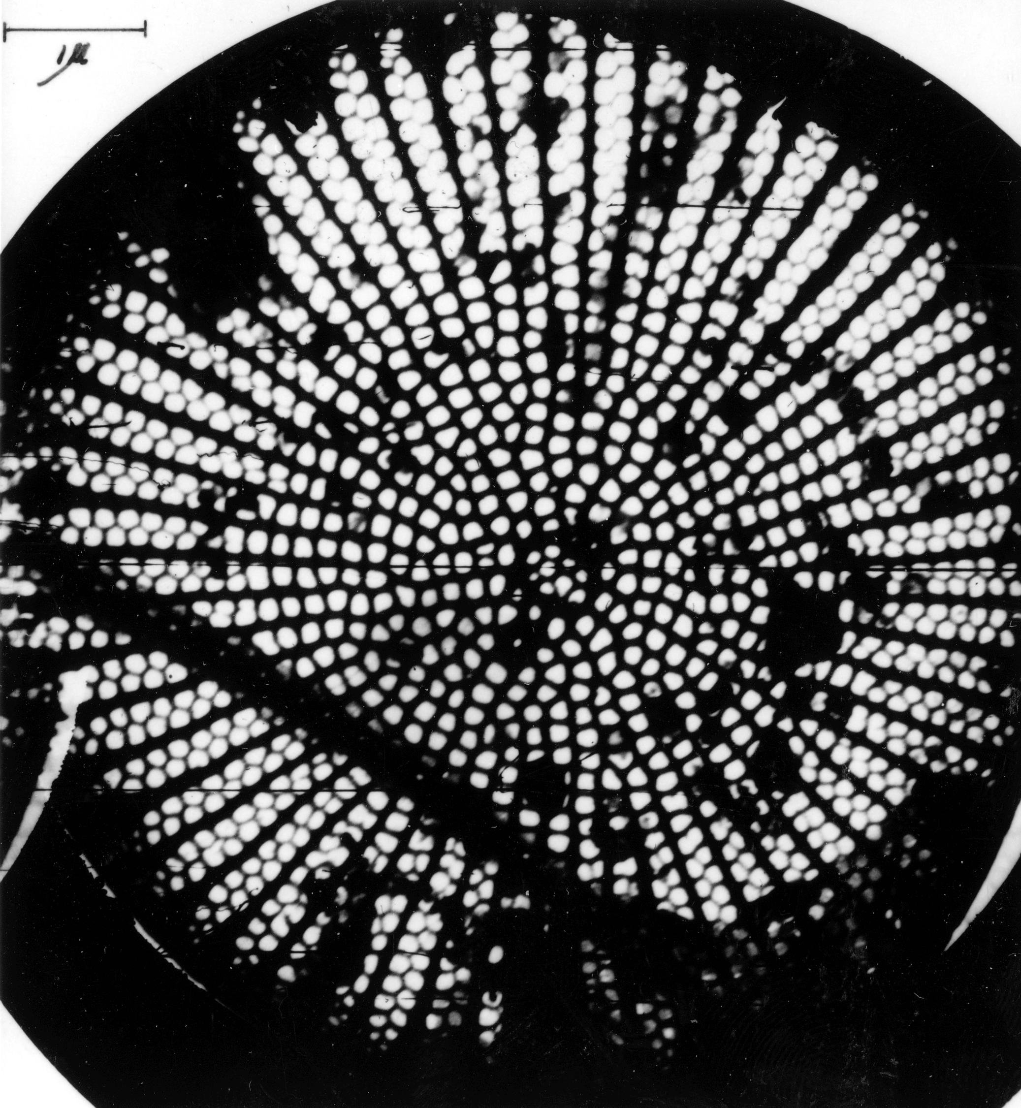
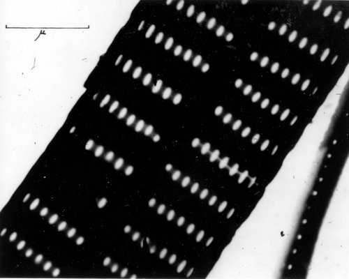
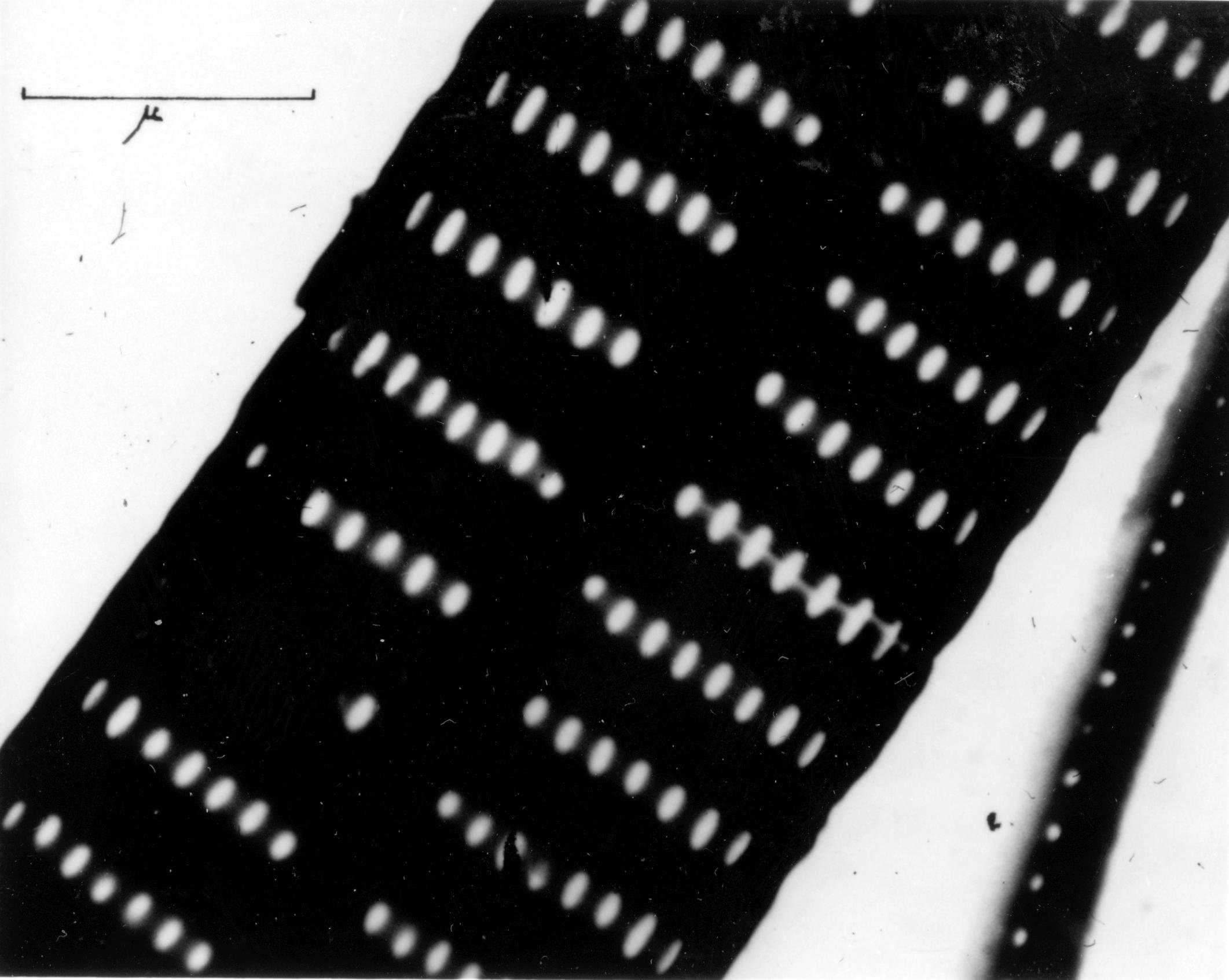
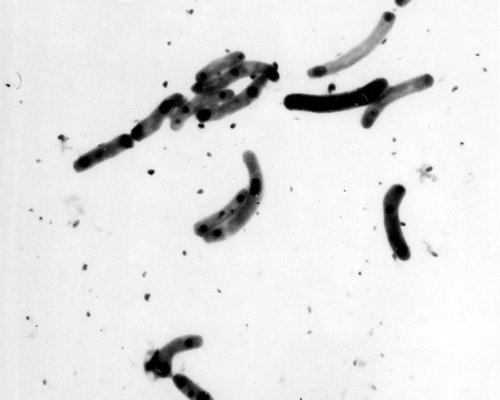
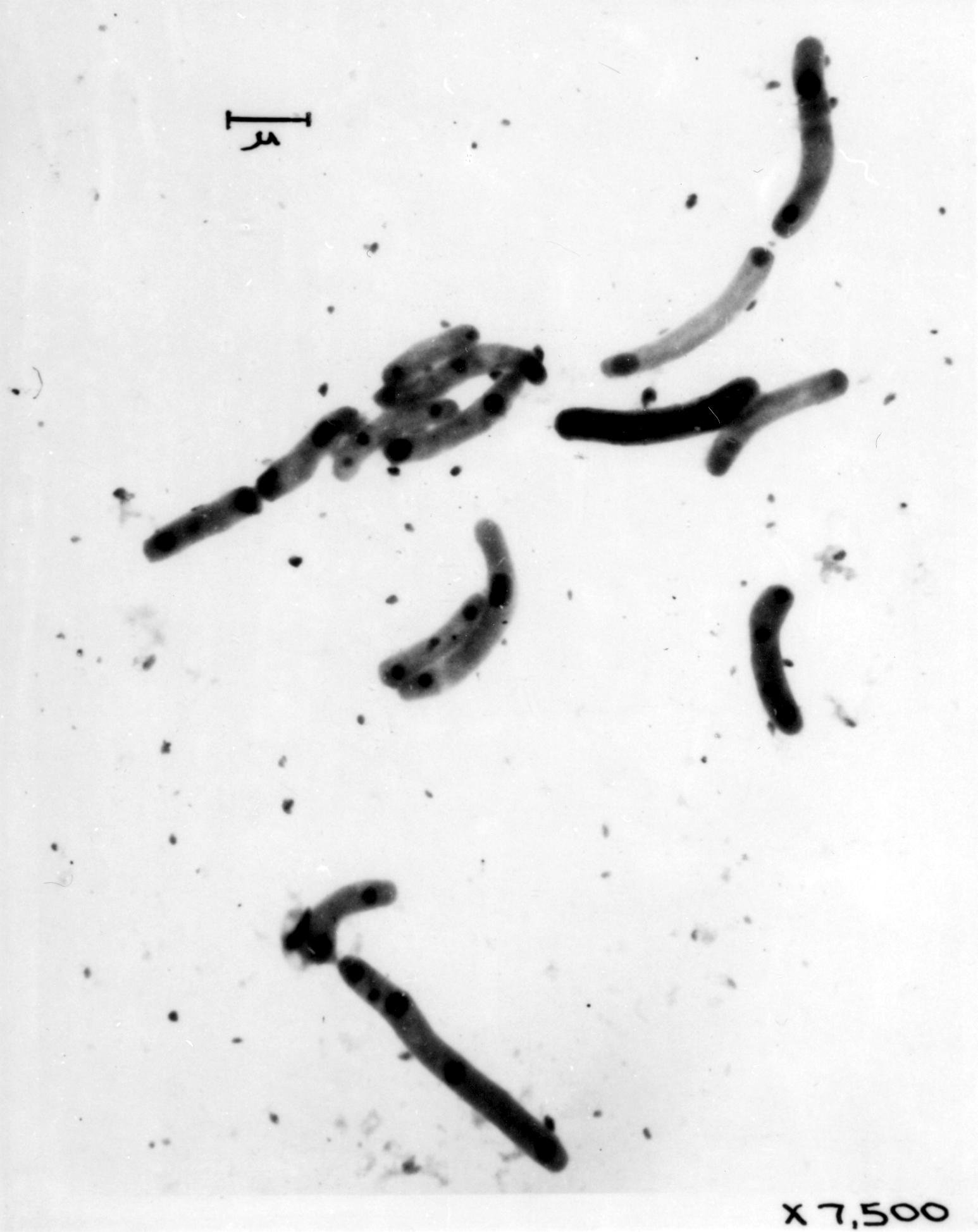
I studied the structure of diatoms with Dr. D.H. Hamly of the Biology Department and published two papers (28, 29) with him on such delicious-sounding diatomic names as "Pleurosigma angulatum," "Synedra delicatissima" and "Amphipleura pellucida" (Fig.'s 7A, 7B). With the 1938 model, and com-pletely inadequate techniques, Dr. Hamly and I attempted to study the structure of stomata in leaves and the wall structure of the aloe. I frequented the Connaught Laboratories on Campus and the Banting Institute down College Street for non-classified specimens of bacteria and viruses (Fig. 8). I micrographed specimens of the tubercle and typhoid bacillus (Fig. 9) and of Rickettsia typhi and of the vole bacillus (Fig. 10). Canada Post issued a 37 cent stamp in 1988 to commemorate the construction of the 1938 microscope. To my surprise, Figure 10 appeared in the background of the stamp taken from my thesis!
Formvar film replicas and shadow casting were used in 1941 in a war-related project to compare the efficiency of fine particle grinding versus flaming for the polishing of glass surfaces. The film replica, formed by dipping the clean glass into an ethylene dichloride solution of Formvar and then drying it, adhered so stubbornly to the clean glass that it would not strip or float away on a water surface. We had the same difficulty whenever we prepared Formvar films for specimen supports. Dr. Burton, passing by and noticing my difficulty would caution me, "tease it, Watson! tease it." Because I was engaged in a stripping operation, this became known around the laboratory (graduate students being how they are), as the "Watson strip-tease technique" of no known relation to the terpsichorean art being practiced in downtown Toronto in those days at the Roxy Theatre.
We did finally discover a method for removing such films, or replicas easily from glass surfaces. It was decided that the glass surfaces were just too clean to be stripped. We needed a technique which would be readily available, would allow easy stripping and would leave a clean replica. To effect this, I learned to draw the "too clean" slide gently and routinely over my nose, (being very careful not to scar that organ). I would then proceed to rub the slide, plus my nose oil vigorously with a clean cloth. By this procedure, a thick film of oil always remained on the slide which was dipped into the Formvar solution. After drying, and with the end and sides of the slide scored, this technique always allowed the film to float away easily and cleanly on a water surface. We never revealed the secret of our success, until now. Indeed, for many years electron microscopy was more an art than a science and many of our methods were similar to those of the early fliers who flew "by the seat of their pants." Much of early electron microscopy was done the same way.
The 1938 microscope was the only such instrument operating in the Allied world during the earliest days of World War II and was one of the very few devoted almost exclusively to war-oriented problems throughout the war years. We who operated it were checked out as A-1 fit by the Army every 6 months and then waited rather expectantly for the notices to arrive requesting our presence elsewhere. The notices never came. We were all sworn to secrecy about what we were doing and we wrote many reports which never saw the light of unclassified day.
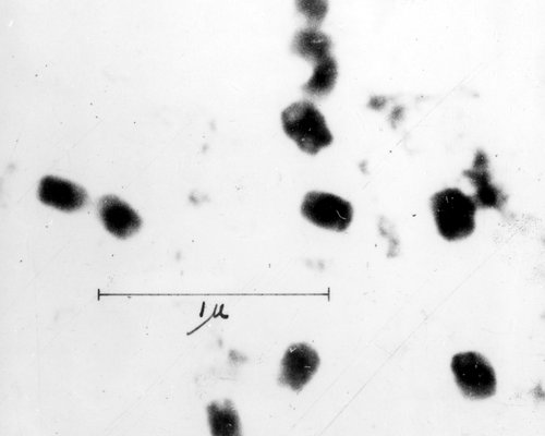
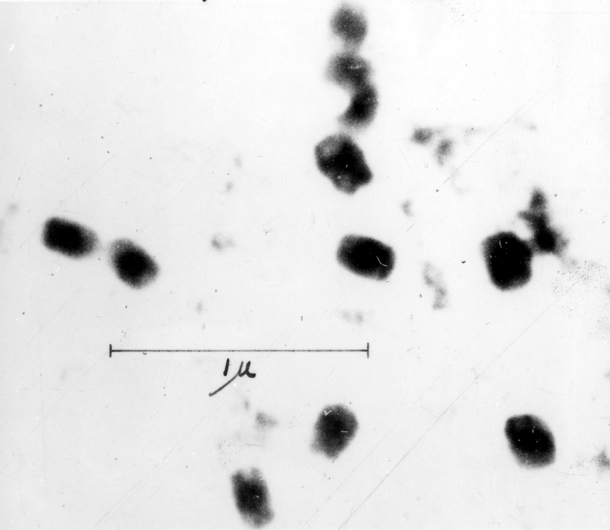
Much of the time of Dr. Newman, Dr. Deacon and myself was given to studies of the particle sizes and other properties of debilitating, solid particle aerosols, as related to gas masks and their construction. Our usual test object was a dispersion of round methylene blue particles (Fig.11), atomized from methylene blue solutions. Many other varieties of aerosols and smokes were also examined, by drawing a known volume of the smoke past many filter types at a range of velocities. The effluent particles were mounted by means of a thermal precipitator, adapted for use with an electron microscope grid. With none of the modern methods of measuring and counting particles available to us, countless hours were spent standing before a ruled, white wall screen, ruler in hand, recording number and sizes of particles from projected micrographs, for our later statistical analysis. In November 1944, the final report on efficiency and particle size was submitted by the three of us with Burton.
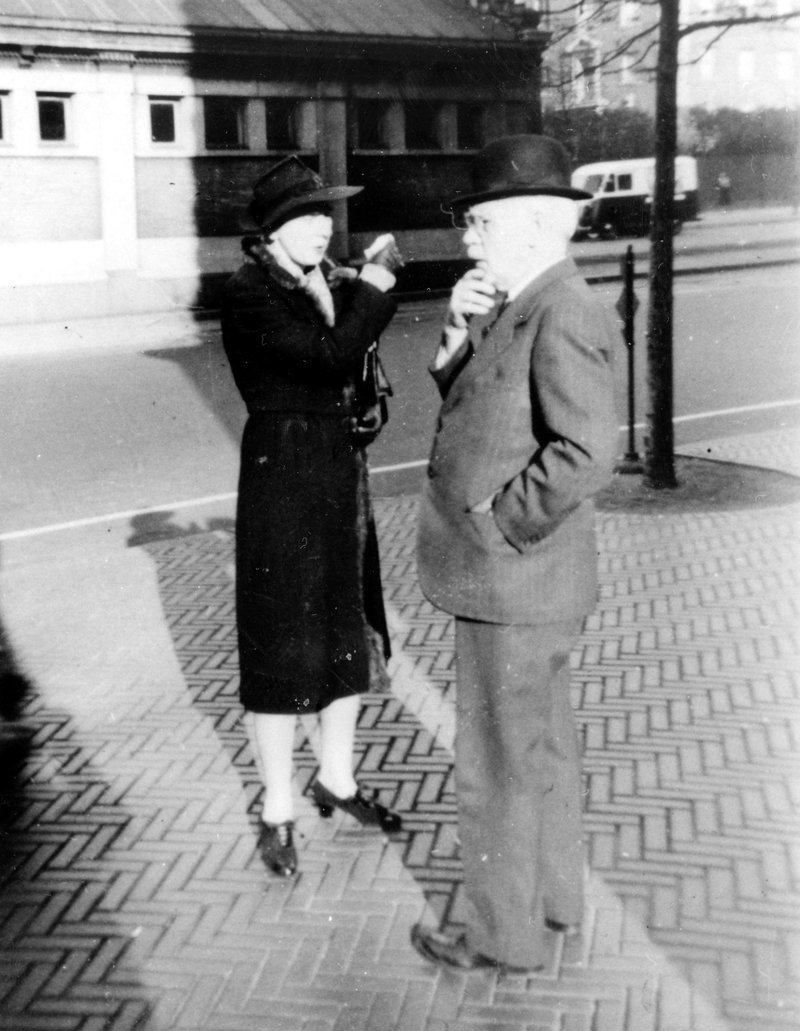
Fig. 14 A puzzled Professor Burton and a helpful Dr. Deacon on a New York City street in 1942, deciding how best to find their way with Dr. Newman and the Author to Columbia University, there to discuss war-related electron microscopy.
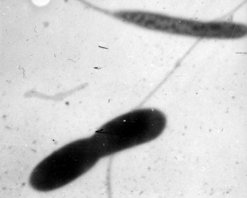
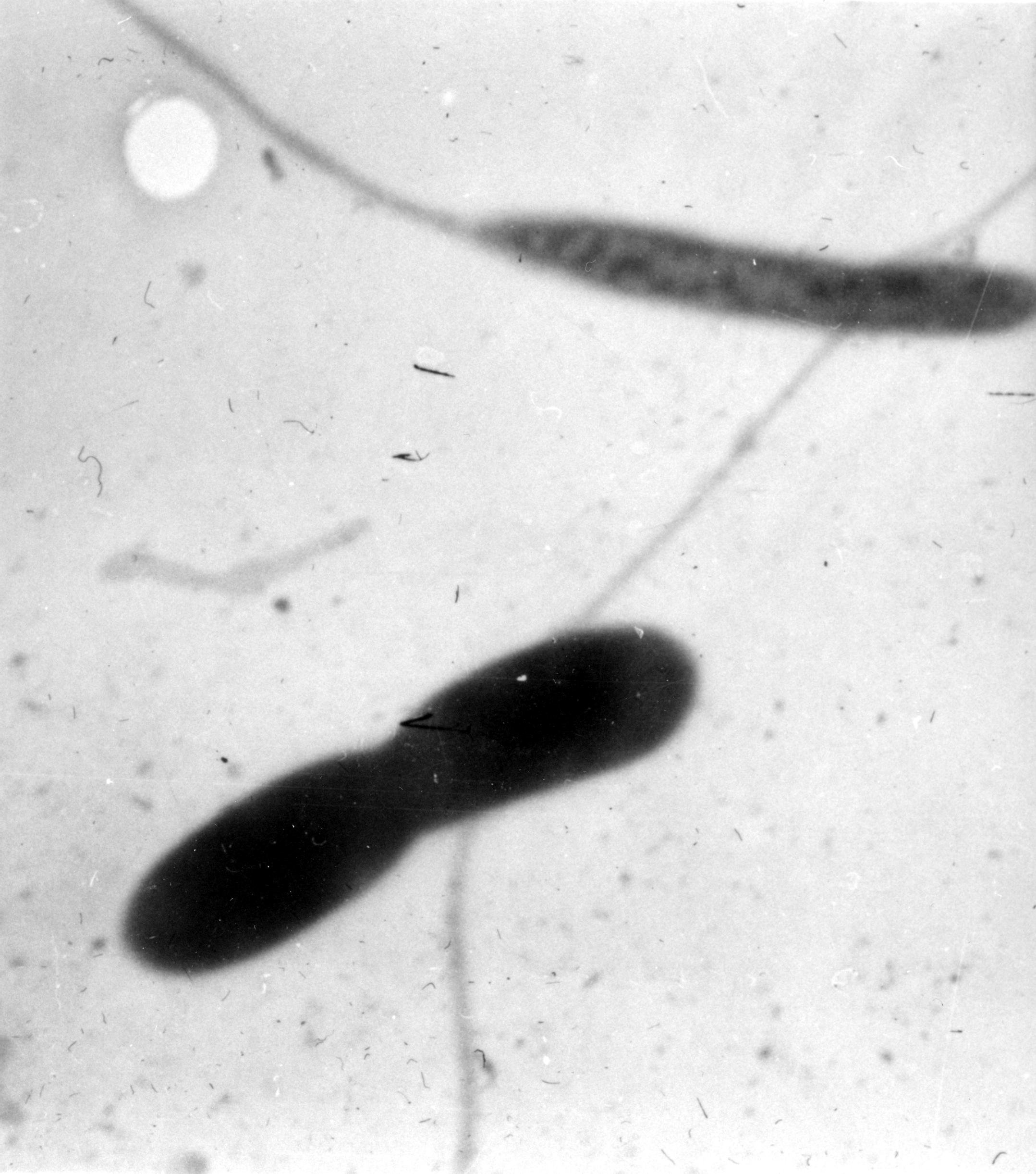
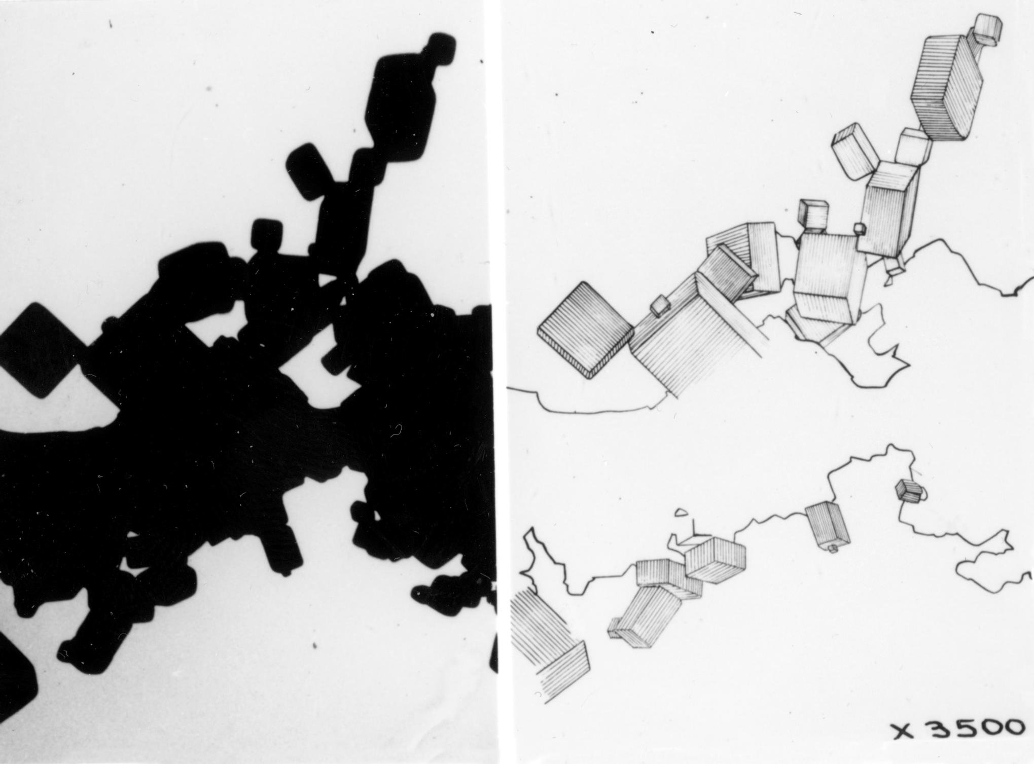
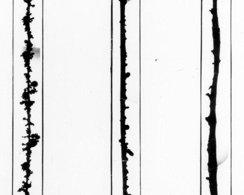
During the war years, Dr. Burton did succeed in publishing one brief paper (30) on electron microscopy, which described the capture of small air-borne particles by single fibers (Fig. 12). In the text, Burton gives the authorship as Burton, Newman, Deacon and Watson, but ever generous, he pleased me much by stating at the end of his article, "this particular experiment was done by the last name." In another similar experiment, I stretched three single fibers of different diameters over the open end of a specimen cartridge with no supporting film or grid, in order to test the theoretical conclusion that the smallest diameter fiber would catch air-borne particles most efficiently when they were drawn past the fibers, (Fig. 13). Particle size investigations were also carried out to determine which size of aerosol particle would be most efficient for light scattering with reference to generating the most opaque smoke screens at sea. In 1942, these investigations saw Burton, Newman, Deacon and myself taking a trip to New York City to discuss light scattering with Columbia University scientists, (Fig. 14).
Looking back on those delightful Toronto days, I wish that there had been someone to encourage us to publish our work. Not only did the University give away its great lead in electron microscopy, but it published very little of the work that was done. At various times during my four years with the Department, a whole smorgasbord of specimens was studied, in my case often in the hope of finding subjects sufficiently unclassified as to be included into my thesis. A variety of polishing powders was examined, demonstrating that their fine structure was observable to a degree not possible with the light microscope. Due to war-time transportation difficulties, we were asked to compare the crystal fine structure of paper-coating clays from English and North American sources. From a combination of electron microscopy and diffraction, it was decided that there was no difference between the flat hexagonal crystals in all specimens, estimated from intensity comparisons to be a minimum of 150 Å thick. Glass and cellulose fibers, asbestos dust and typhoid bacteria in the presence of bacteriophage were examined, as was the effect of growing Streptococcus hemolyticus in the presence of sulpahthiosole. None of these investigations were ever published, except partly in my thesis and in Burton and Kohl's early book (17). Fig. 15 shows fibers of cuprene, a polymer of acetylene formed in the presence of copper and Fig. 16 is a field of soap particles in a sample of lubricating grease spread out on a collodion film. Methods of stereo-electron microscopy were also developed with the 1938 instrument. Indeed, a lecture given in 1942 on this subject and a paper published in 1943 (25) resulted in my first job offer, following the Ph.D.
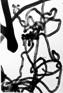
Fig. 15 Micrograph of Cuprene fibers, magnification unknown.
Silicosis was a major problem in those days among Ontario miners, particularly for those who engaged in drilling operations. Dr. Burton was anxious to learn whether the electron microscope could contribute to a better under-standing of the disease. The particulates in the atmosphere of the mine were collected on electron microscope specimen grids, using the adapted thermal precipitator in the mine, and the properties of the dust created by drilling were compared with those created by blasting operations. It was eventually determined that drilling produced much more of the smaller and medium-sized dust particles than did the blasting. It was theorized that the medium-sized particles probably remained longer in the lungs than either the very small or the very large particles, and would be most liable to account for the increased virulence of the disease among drilling miners. In 1942 an apparently important project came our way, a study of certain thin metal screens which were received periodically from the National Research Council and concerning which reports of our work were requested with some urgency. We were to take micrographs of holes in the screens, determine their average size and range, and measure the mean free space over the screens. We were not told the purpose of the exercise.
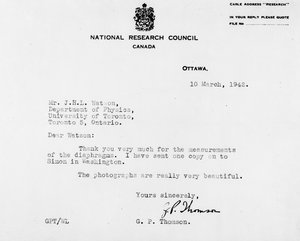
Fig. 17 A letter received by the Author in 1942 from G.P. Thomson.
Although we examined many screens and wrote many reports, we heard nothing about them except for a treasured letter (Fig. 17) I received in 1942 from Dr. G.P. Thomson, physicist, co-discoverer of electron diffraction (Davisson and Germer discovered it separately), and the son of Sir J.J. Thomson who discovered the electron. At first the openings in the screens were long and narrow, rough-edged and quite large, scarcely needing an electron microscope to see them. Gradually, as fresh screens arrived, the openings became sub-microscopic, much smoother in outline and more uniform. Dr. Burton seemed to know as little about the nature of the work as we did and as we grew bored with it, he would encourage us to examine other specimens, fly's wings for example. Invariably, as we relaxed on the project, we would have a "wakeup" meeting with Dr. Thomson and a Dr. Simon, who would visit the laboratory. I can still see them, in my mind's eye, standing over in a corner out of ear-shot, discussing our micrographs and our results. Following those visits, we would find ourselves back on the screens! It wasn't until World War II was long over, while reading an unclassified report concerning the Manhattan Project that I noted Thomson's and Simon's names prominently displayed and realized that our work with the screens might have had more significance than we knew, perhaps for a separation of isotopes by gas diffusion through submicroscopic holes.
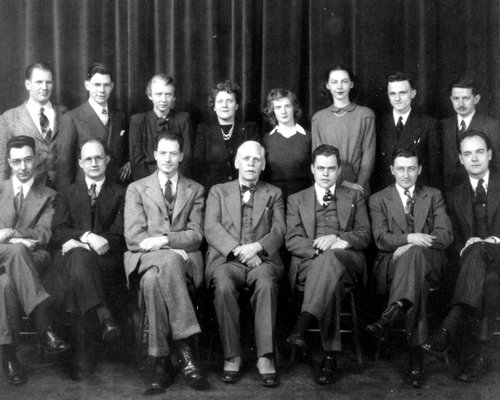
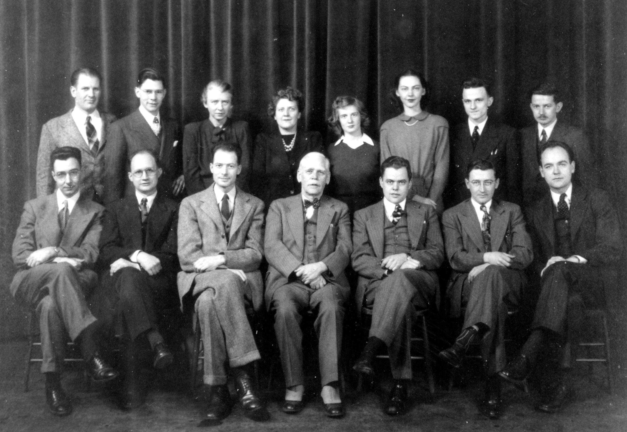
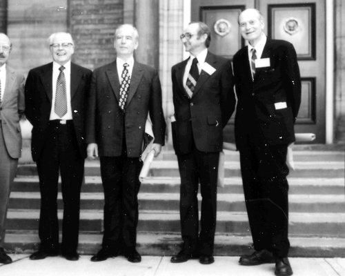
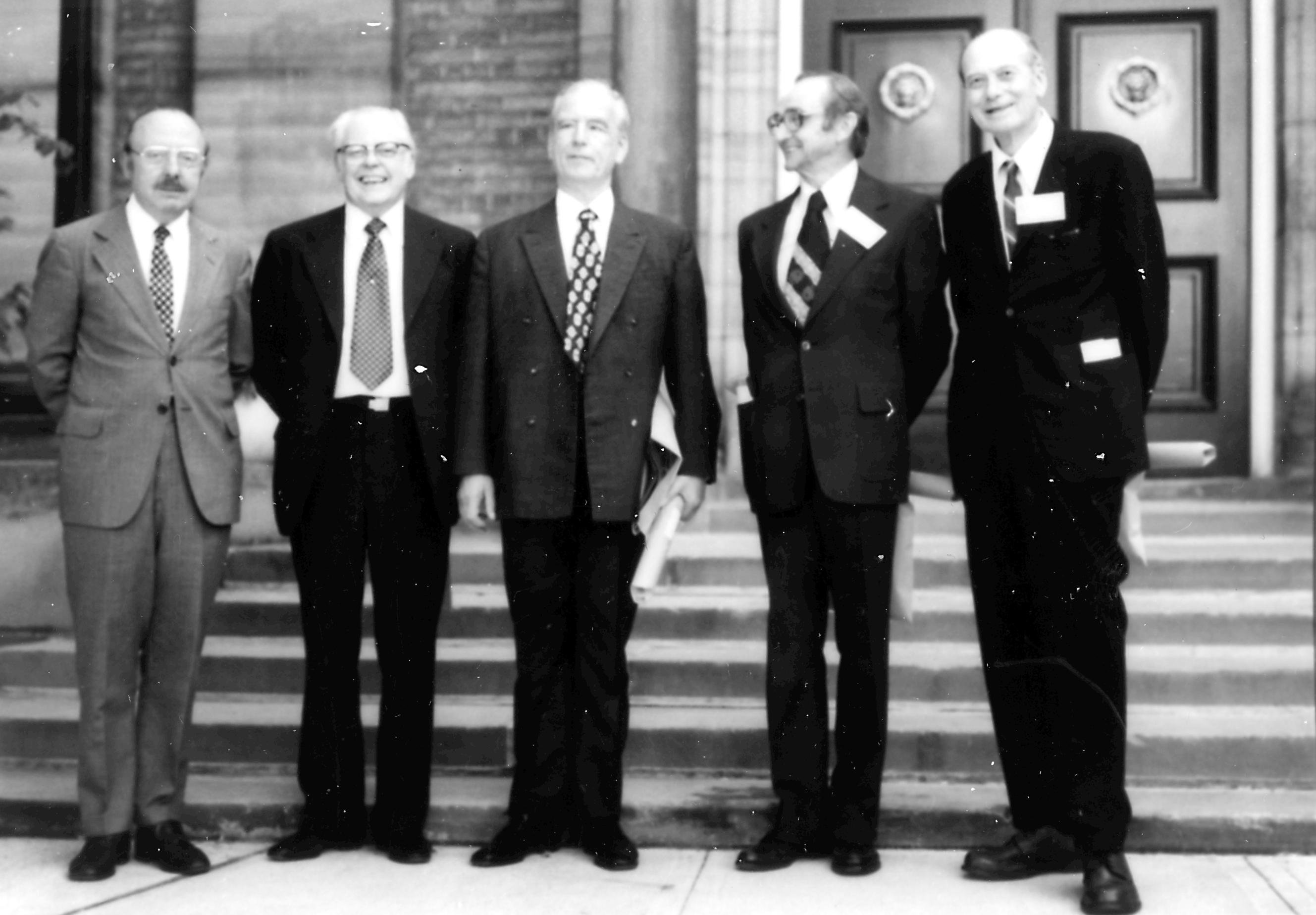
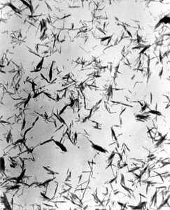
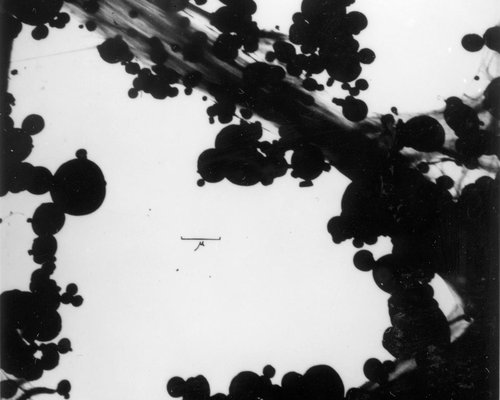
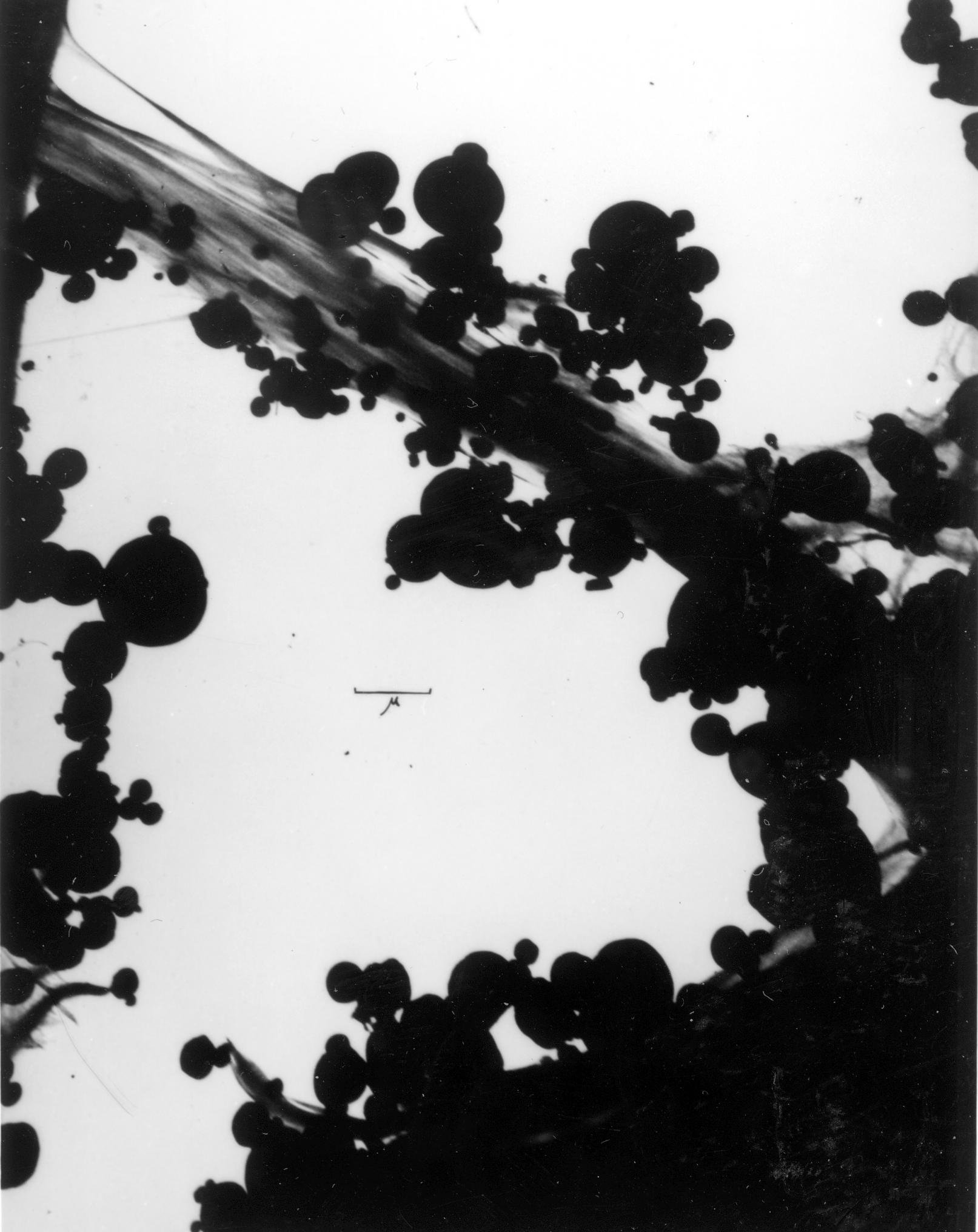
Fig. 16: Micrograph of soap particles in a sample of lubricating grease, spread on a collodion film, X.
It was during those very early days that Alice Grey was bringing over large chunks of pathological human tissue from Banting Institute for electron micro-scopic examination. These chunks were macerated in a blender in the vain hope that a few areas of sufficient thinness, to allow electron penetration, might be found to yield useful information. To fastidious physicists, used to somewhat drier fields, this procedure was far from elegant! By hindsight, it can be seen that the attempt was but a first faltering step in the long process of developing thin section specimen preparation techniques for electron microscopy. Memories of Professor Burton bring back to me many events which are important to electron microscopy. For example, he would frequently ask us to micrograph an identical same field by both electron and optical microscopes, in order, of course, to demonstrate the superiority of the former, never minding that the conditions for the one were not liable to be at all ideal for the other instrument. Again, by hindsight it can be seen that Burton was as usual not far off the mark. It is still a good idea to couple light and electron microscopy. Today, with the availability of so much new instrumentation and techniques for microscopy, it is even more desirable to couple many instruments in the solution of microscopy problems. Regardless of resolution and magnification, the characteristics of any sample as seen by visible light as a consequence of its absorptive properties for light are not necessarily the same as the same sample's characteristics as "seen" by electrons which depend upon the electron-scattering properties of the specimen. Each instrument produces its own image and each is important in interpretation.Glen Ellis, who came to the laboratory about the time I was leaving it temporarily, told me that one day in 1945 when he was casting about for a suitably thick specimen with which to test the performance of apertures in the 1944 instrument, an obliging fly flew in an open window and offered one of its wings for the purpose. Dr. Burton was so intrigued by the resultant micrographs that he sent biologist, Mary Ferguson, to the Dominion Fly Farm to collect fly's wings from numerous specimens. Doing her M.A. thesis, Mary so clogged the works with flies wings that it is said there were more flies in the Department of Physics than there were in Yankee Stadium, or so Ellis reported! But, that was Professor Burton, always enthusiastic and a real fan of electron microscopy. Professor Burton attended and gave a brief paper at the 6th meeting of the Electron Microscopy Society of America held in Princeton in 1948 on "Micrographs of Insect Wings." He died in July of 1948. It is regrettable that he did not live to see more of how his original faith in the electron microscope was to be vindicated in the years that have followed. In September of 1948 the E.M.S.A. held its annual meeting at the Department of Physics at the University of Toronto. The meeting was dedicated to his memory, and in recognition of his pioneer work in introducing electron microscopy to the Western Hemisphere, the first annual award in his name was made to a promising young scientist in the field, the EMSA Burton Medal.Figure 18 shows the faces of many of those early Canadian electron microscope people, as they were, during a conference hosted by Professor Burton at the McLennan Laboratory in 1947. Only Albert Prebus was missing. Fig. 19 was taken near Convocation Hall in August 1978, just after honourary degrees had been conferred by the University on five men who had pioneered so much in electron microscopy.
I do not know anymore than I have already recorded here, concerning the people and events involved in electron microscopy at Toronto after 1944. I have referred to Ellis who received his Ph.D. in 1946 and to some of the work done by Boswell who won his Ph.D. in 1950. Electron microscopy continued to be applied in the Department of Physics into the early 1950's, involving, the late Dr. David Scott with Tom McLaughlan and R.S. Sennett, as well as Frank Boswell. I know that research was in progress on thin films as well as microcrystals but I am not familiar with the details and so must leave any report of these later years to those who are familiar with them. I have described the final resting place of the 1938 instrument and I have been told that the 1940 model is in a museum in Washington. The whereabouts of the 1944 model is less certain but I have again been told that it rests in someone's basement!The very earliest Canadian electron microscopists, Burton, Kohl, Hall, Hillier, Prebus, Watson, Ladd, Newman and Deacon mastered the electron microscopy of the period and gathered basic knowledge of many subjects which even experts in those subjects did not and could not know about the ultrafine structure of pigments, colloids, aerosols, ceramics, metals, greases, oils, viruses, bacteria, microcrystals whatever was examined with the Toronto electron microscope of 1938. We studied them all with the electron microscope and were the first to do so.During the summer of 1944 (Fig. 6) when I had returned to the laboratory to help bring the third microscope into operation, I remember that at some point, my father had suggested that he and I should go north for a week's trout fishing. Asking for a week off, Dr. Burton exploded, "four years of work and just as the instrument is to be turned on you go trout fishing?" - I went fishing. What wonderful years they were.
Figure Captions and References
Captions
Fig .1A The 1938 Toronto model electron microscope, as set up originally, with the single plate or roll-film camera. Parts of the high tension, vacuum system and lens current control rheostats are shown.
Fig. 1B The 1938 column with the revised camera and its cross-sections.
Fig. 1C Prebus left and Hillier right, working with the microscope in 1938.
Fig. 1D The 1938 microscope with the revised camera, Prebus at the microscope, Professor Burton left and Ladd sitting right, 1940.
Fig. 2 Canadians interested in electron microscopy met in the Spring of 1947 at the Shawinigan Chemicals Co. in Shawinigan Falls, Quebec. Glen Ellis is at far left in the front row. Dr. Keyes of McGill University is beside him and Professor Burton and James Hillier are beside Keyes in the front row, with the Author's head between them in the second row. Others are representatives from McGill University, the National Research Council and the staff of the Shawinigan Company.
Fig. 3 Micrograph of pneumoccus, taken very early by Hillier. X
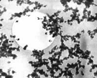
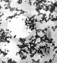
Fig. 4 Micrograph of a dispersion of carbon black particles, deposited from a methane flame upon a collodion supporting film. X. A very acceptable compensation of the objective lens is suggested by the symmetrical appearance around the hole in the film.
Fig. 5 The column of the 1944 Toronto microscope set up on a laboratory table and the cross-section of its column is shown. The instrument is almost complete but not yet operating.
Fig. 6 A young Watson pondering a problem with the specimen cartridge of the 1944 model in the summer of 1944.
Fig. 7A. Micrograph of a diatom of the Cyclotella species, X.
Fig. 7B Micrograph of part of the skeleton of the diatom Synedra delicatissima, X.
Fig. 8 Micrograph of vacinia viruses, X.
Fig. 9 Micrograph of typhoid bacillus, with flagellae, X.
Fig. 10 Micrograph of vole bacilli, X, which resemble the tubercle bacillus. We were comparing their morpholo-gies. Polar bodies are seen.
Fig. 11 Micrograph of methylene blue particles caught on fibers of rice lens paper, with no supporting film, X.
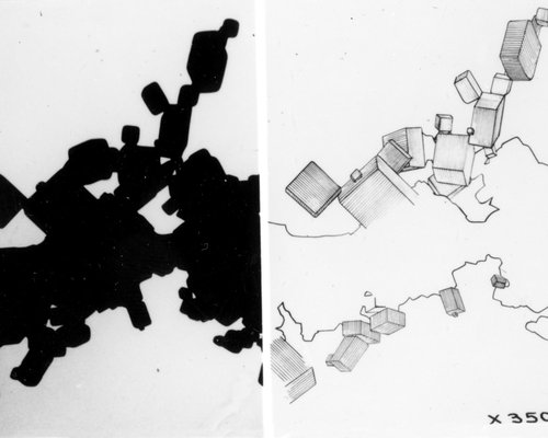
Fig. 12 Micrograph and schematic of tiny sodium chloride crystals caught from an air stream by a single fiber, X.
Fig. 13 Micrograph at low magnification, of methylene blue particles caught from an air stream upon three fibers of different diameters to demonstrate the increased catching efficiency of the smallest diameter fiber, approx. X500.
Fig. 14 A puzzled Professor Burton and a helpful Dr. Deacon on a New York City street in 1942, deciding how best to find their way with Dr. Newman and the Author to Columbia University, there to discuss war-related electron microscopy.
Fig. 15 Micrograph of Cuprene fibers, magnification unknown.
Fig. 16 Micrograph of soap particles in a sample of lubricating grease, spread on a collodion film, X.
Fig. 17 A letter received by the Author in 1942 from G.P. Thomson.
Fig. 18 Those attending a conference at the University of Toronto in 1947, hosted by Professor Burton; front row (l. to r.) Lorne Newman, Chester Calbick, Cecil Hall, Professor Burton, James Hillier, Bill Ladd, John Watson; back row (l. to r.) Tom McLaughlan, Frank Boswell, Beatrice Deacon, Alice Grey, Mary Ferguson, Barbara Woods, Roy Sennett, Glen Ellis.
Fig. 19 Honourary Graduands, after a special summer convocation in 1978 held by the University of Toronto, during the annual meetings of the international Federation of Electron Microscopy Societies, hosted by the Canadian Microscopical Society, (l. to r.) Keith Porter, James Hillier, Ernst Ruska, Albert Prebus and Cecil Hall.
References
- Franklin, U.M., Weatherly, G.C., Simon, G.T., "A History of the First North American Electron Microscope," Ninth International Congress on Electron Microscopy, (Ed. J.M. Sturgess), III 3-25 (1978)
- Hall, C.E., "Recollections from Early Years: Canada - U.S.A.," from The Beginnings of Electron Microscopy, Ed., P.W. Hawkes, 275-296, Academic Press, London (1985).
- Doane, F.W., Simon, G.I., Watson, H.H.L., "An Historical Account of the Development of the First Electron Microscope in North America," The Microscopical Society of Canada, 20th annual meeting, Toronto (1993).
- Watson, J.H.L., "Microscopy and Music - Dimly Related," New York Microscopical Society Year Book, 10: 51-64 (1964-1965).
- Watson, J.H.L., "Toronto Daze," Bull. Micros. Soc. Can 2 (4): 10-18 (1974).
- Watson, J.H.L., "The Origins of Electron Microscopy on the North American Continent at the University of Toronto - A Recollection," E.M.S.A. Bull., 22 (2): 97-106 (1992)
- Watson, J.H.L., "The Origins of Electron Microscopy on the North American Continent at the University of Toronto - Part II," M.S.A. Bull., 23 (2): 139-145 (1993).
- Watson, J.HL, "Very Early Canadian Electron Microscopists and Their Microscopy," Micron (in press), Proc. 22nd annual meeting of Microscopical Society of Canada, Ottawa (1995).
- Hillier, J., "The Fundamental Theory of Electron Optics and its Application in the Construction of a New Magnetic Electron Microscope," M.A. Thesis, University of Toronto (1936).
- Hall, C.E., "An Electrostatic Electron Microscope," McGraw-Hill, N.Y., 1st Ed., 1953; 2nd Ed. 1966.
- Hall, C.E., Introduction to Electron Microscopy," McGraw-Hill, N.Y., 1st Ed., 1953; 2nd Ed. 1966.
- Prebus, A., "Improved Pole-piece Construction of the Objective Lens of a Magnetic Electron Microscope," Can. J. Res., A-18: 175-177 (1940).
- Prebus, A. and Hillier, J., Can. J. Res. A-17: 49-63 (1939).
- Burton, E.F., Hillier, J., Prebus, A., "Report on the Development of the Electron Supermicroscope at Toronto," Phys. Rev., 56:1171-2 (December 1, 1939).
- Hillier, J., "The Effect of Chromatic Aberration on Electron Microscope Images", Can. J. Res., A-17:64-69 (1939).
- Burton, E.F., Hillier, J., Prebus, A., "The Contribution of the Electron Microscope to Medicine. 1. The Electron Microscope Described," Can. Med. Assoc. J., 42:116-119 (1940).
- Burton, E.F. and Kohl, W.H., "The Electron Microscope," Reinhold Publishing Corporation, New York, 1st Ed. (1942), 2nd Ed. (1946).
- Ladd, W.A. "The Magnetic Electron Microscope and its Applications," M.A. Thesis, University of Toronto (1940).
- Watson, J.H.L., "The Measurement of the Magnetic Field Along the Axis of a Magnetic Electron Lens," M.A. Thesis, University of Toronto (1940).
20. Watson, J.H.L. "Electron Microscopy," Ph.D. Thesis, University of Toronto (1943).
21. Hillier, J., "The Fundamental Principles of Practical electron Microscopy," Ph.D. Thesis, University of Toronto (1941).
22. Prebus, A., "The Construction of a Magnetic Electron Microscope of High Resolving Power," Ph.D. Thesis, University of Toronto (1940).
23. Watson, J.H.L., "An Effect of Electron Bombardment Upon Carbon Black," J. Appl., Phys., 18:153-161 (1947).
24. Watson, J.H.L., "Specimen Contamination in Electron Microscopes," J. App. Phys., 19:110-111 (1948).
25. Hillier, J. "The Fundamental Principles of Practical Electron Microscope of High Resolving Power," Ph.D. Thesis, University of Toronto 1940).
26. Boswell, F.W.C., "Precise Determination of Lattice Constants by Electron Diffraction and Variations in the Lattice Constants of Very Small Crystallites," Proc. Phys. Soc. Amer., LXIV: 465-475 (1951).
27. Boswell, F.W.C., "Electron microscope Studies of Virus Elementary Bodies," Brit. J. Exper. Path., XXVII:253-260 (1947)
28. Hamly, D.H. Watson, J.H.L., "Potentialities of Optical and Electron Microscopes," Trans. Roy. Soc. Can., 35:61-65 (1941).
29. Hamly, D.H., Watson, J.H.L., "Electron and Optical Microscope Interpretation of the Wall of Pleurosigma angulatum," J. Optic. Soc. Amer., 32:433-445 (1942).
30. Burton, E.F., "Electron Microscope Studies of the Capture of Air-borne Particles by Single Fibers," Nature, 152:540(1943).